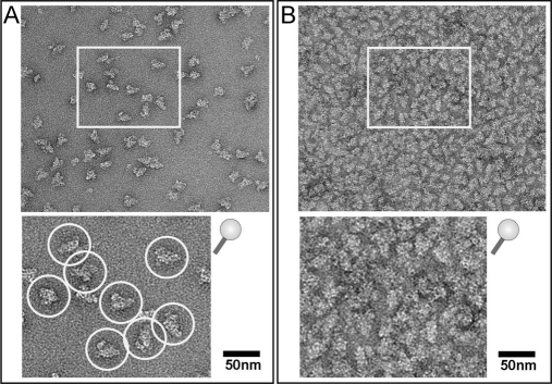
Fig. 4.
Electron microscopic analysis of human U4/U6.U5 tri-snRNP complexes. A, negatively stained EM image of ∼5 fmol of tri-snRNP per specimen. Individual particles showing the typical triangular shape of tri-snRNP can be distinguished (upper panel, overview image; bottom panel, subwindow at a higher magnification as indicated in the upper panel). Such a particle density is suitable for single particle image processing. Carbon films with adsorbed particles of this density were subjected to ECAD analysis. B, negatively stained EM image of tri-snRNP showing a particle density too high for image processing; exact counting of the particles is not possible at this high density (upper panel, overview image; bottom panel, subwindow at a higher magnification as indicated in the upper panel). Such a particle density is unsuitable for single particle image processing. These overloaded carbon films can in principle be analyzed by ECAD to increase the sample amount subjected to digestion; however, such overloading is not suitable for a correlation of the MS and EM analyses.

