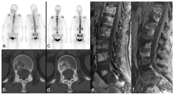
Figure 9.
Scintigraphic flare. (a) Numerous bone metastases show tracer uptake on a Tc 99m MDP bone scan in a patient with breast cancer. (b) Companion CT examination demonstrates a lytic metastasis in the L1 vertebral body. (c) Six months later, the lesions demonstrate increased tracer uptake. (d) Companion CT shows sclerotic fill-in of the lytic lesion, which can occur with disease progression or healing. (e, f) Fat-saturated T1-weighted sagittal MRI examinations of the lumbar spine obtained (e) 1 month and (f) 2 months after the bone scans show a decrease in the size and/or enhancement of the metastases, indicating a positive response to therapy. Incidental note is made of interval insufficiency fracture of the superior endplate of L4 on (f). The increased MDP uptake on the bone scan (b) was the result of healing sclerosis and representative of a scintigraphic flare in a patient undergoing partial response rather than progressive disease.

