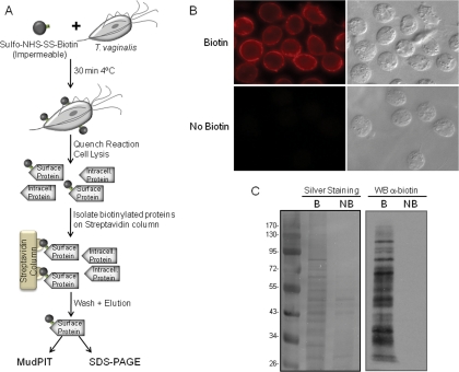
Fig. 1.
Biotinylation of T. vaginalis surface proteins using sulfo-NHS-SS-biotin. A, scheme of the protocol for biotinylating and purifying surface proteins. B, immunofluorescence assay with anti-biotin antibody. Top panel, biotinylated parasites; bottom panel, non-biotinylated parasites. Data for only one strain (T1) are shown; however, all had comparable staining patterns. C, SDS-PAGE analysis of proteins purified by streptavidin. Biotin-labeled (B) and unlabeled proteins (NB) were recovered by affinity purification, separated by SDS-PAGE, and silver-stained (left panel) or detected by Western blot (WB) with anti-biotin antibody (right panel). The molecular weight marker is shown on the far left. Intracell., intracellular.

