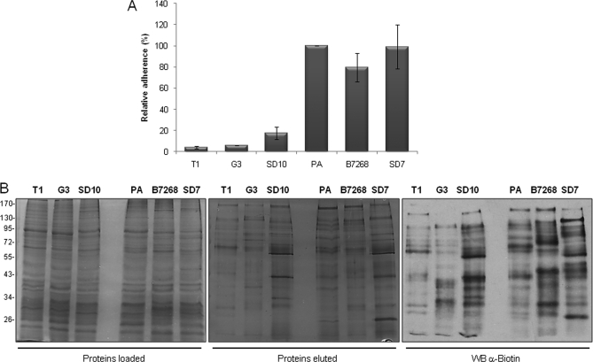
Fig. 4.
Analyses of six parasite strains for adherence and biotinylation. A, binding to ectocervical cells. Parasites from six different strains were fluorescently labeled and incubated with ectocervical cell monolayers at 37 °C. Coverslips were washed to remove non-adherent parasites, mounted, and visualized and quantified by fluorescence microscopy. Data are expressed as percentage of adhesion related to the PA strain ± the standard deviation of the mean. A representative experiment of three independent experiments is shown. B, surface protein biotinylation of strains with different adherence capacities. A comparable amount of protein extract of three less adherent (T1, G3, and SD10) and three highly adherent strains (PA, B7268, and SD7) was loaded onto a streptavidin column (left panel). Biotinylated labeled proteins were recovered, separated by SDS-PAGE, and silver-stained (middle panel) or detected by Western blot (WB) with an anti-biotin antibody (right panel). The molecular weight marker is shown on the far left.

