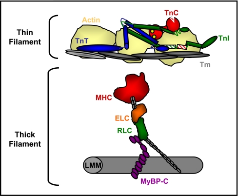
Fig. 1.
Schematic illustrating cardiac sarcomeric proteins. The myofibrillar lattice is composed mainly of actin thin filaments and myosin thick filaments, each bound to regulatory proteins. Activation of cardiac contraction during systole proceeds by calcium binding to troponin C (TnC), which induces conformational changes and altered interactions among troponin I (TnI), troponin T (TnT), and tropomyosin (Tm), resulting in the removal of steric inhibition over the myosin binding site on actin. An activated thin filament allows the binding of myosin heads, which then propel the actin filaments toward the center of the sarcomere. The thick filament is composed mainly of MHC, which binds two light chains, ELC and RLC, and associates with MyBP-C in the hinge and light meromyosin (LMM) regions.

