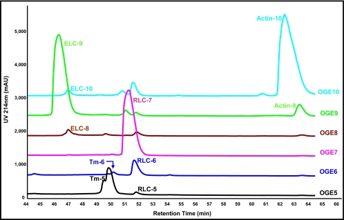
Fig. 4.
Purification of sarcomeric proteins in OGE fractions using RP-HPLC. Shown are OGE fractions 5–10, each run separately on a 4.6 × 150-mm C4 reverse-phase column (TP5415, Grace Vydac) with UV detection at 214 nm, and the chromatograms were overlaid for comparison. Separation of endogenous charge variants of ELC, tropomyosin (Tm), RLC, and actin is demonstrated in the retention time range 44–66 min. Quantification of the three unique RLC species demonstrated a 3.1/18.1/78.8% distribution of endogenous RLC in fractions RLC-5, -6, and -7, respectively, which were collected for further analysis by LC/MS/MS.

