Fig. 7.
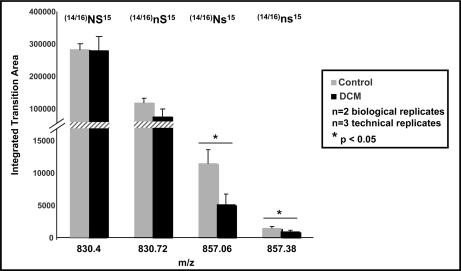
Quantification of human RLC species in DCM versus control by multiple reaction monitoring reveals differences in phosphorylation and deamidation. Heart tissue from non-failing and DCM patients was extracted and fractionated using OGE. Fractions (OGE-7, OGE-8, and OGE-9) from two biological replicates were pooled, and RLC species were separated via RP-HPLC as described above. Fractions were digested using Lys-C and subjected to liquid chromatography tandem mass spectrometry using a triple quadrupole mass spectrometer. The following transitions were monitored for the peptide (K↓RAGGANSNVFSMFEQTQIQEFK↓E) and used to distinguish isoforms: unmodified ((14/16)NS15), 830.4 → 827.4; singly deamidated ((14/16)nS15), 830.70→ 828.4; singly phosphorylated ((14/16)Ns15), 857.1 → 907.4; and singly deamidated/singly phosphorylated ((14/16)ns15), 857.4 → 908.4. Each transition corresponds to the resulting b9 product ion. Individual fractions were analyzed, the area was determined for each transition, and the total area for each transition was combined between fractions for simplicity. Three experiments were conducted, and the averages are shown. Error bars represent the standard deviation. Transitions corresponding to the singly phosphorylated and the phosphorylated/deamidated forms are significantly different between heart disease and control, whereas singly deamidated and unphosphorylated showed no difference.

