Fig. 4.
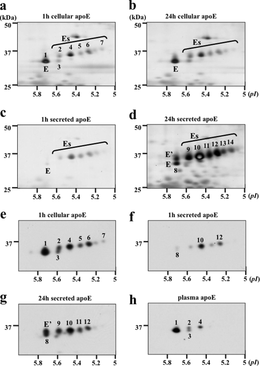
Two-dimensional gel electrophoresis of apoE glycoforms in HMDMs. HMDMs from two independent donors (a–d, donor 1; e–h, donor 2) were cholesterol-enriched by incubation for 2 days with 50 μg/ml AcLDL, washed, and incubated in serum-free and BSA-free RPMI 1640 medium for up to 24 h. At the indicated times, apoE was immunoprecipitated from cell lysates (a, b, and e) and media (c, d, f, and g), separated by 2-DE, and detected by silver staining (a–d) or Western blot (e–h). The distribution of cellular apoE glycoforms did not change between 1 (a) and 24 h (b), whereas there was an increase in the abundance of glycoforms E and E′ relative to Es glycoforms between 1 and 24 h in the medium (c versus d and f versus g). Western blots show that plasma apoE (h) comprises less charged apoE glycoforms than secreted (g) or cellular apoE (e) from the same donor. On the basis of mass and pI, the following glycoforms appear to be the same: 1, E, and 8; 2 and 9; 4 and 10; 5 and 11; 6 and 12; and 7 and 14.

