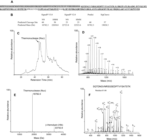
Fig. 1.
Experimental strategy for identification of Nuc in Trap 7. A, combined sequence coverage map of Nuc from trypsin and Glu-C digestion. The underlined amino acids were identified. The shaded region corresponds to the signal sequence. B, signal peptide cleavage site predictions and corresponding predicted masses for Nuc. C, total ion chromatogram of Trap 7 containing a peak corresponding to Nuc. D, raw spectrum of Nuc showing the charge state distribution. Asterisks show charge state distribution of Hlb. E, deconvoluted mass spectrum. F, MS/MS spectrum of N-terminal peptide SQTDNGVNRSGSEDPTVYSATSTK of mature Nuc.

