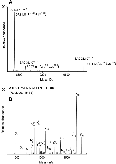
Fig. 9.
A, deconvoluted mass spectrum showing the three forms of SACOL1071 that formed as a result of signal peptide processing at different sites: SACOL10711, signal peptide cleavage at position 26; SACOL10712, signal peptide cleavage at position 26; and SACOL10713, signal peptide cleavage at position 14. B, LTQ-FT-MS/MS spectrum of N-terminal peptide of SACOL10713.

