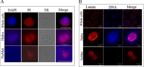
Fig. 2.
RNA and lamin are components of S2 cell NuMat. A, DAPI and PI are seen staining the entire nuclear volume in the whole cell, whereas DNA is seen around the nucleus after extraction in halos, and DAPI signal is lost in the NuMat, indicating complete chromatin digestion. PI staining, which stains both DNA and RNA, persists in NuMat, indicating the presence of RNA component. TR is the transmission microscopic image. B, lamin shows nuclear rim staining in the whole cell, whereas in halos and NuMat the internal meshwork of lamin is accessible for staining. The absence of DAPI signal in NuMat indicates efficient removal of chromatin, leaving clean NuMat in the preparation. Scale bars, 5 micron.

