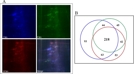
Fig. 5.
Developmental dynamics of NuMat proteome. A, 2-, 8-, and 16-h-old Drosophila embryos were used for NuMat isolation and were separated by two-dimensional DIGE. The NuMat proteome is dynamic during the development of the embryo. B, Venn diagram to show the distribution of spots in various developmental stages of Drosophila embryos. Up to several hundred protein spots can be detected by DIGE, and there are more NuMat proteins in the early stages of development. Colors of the circles correspond to the spot colors in the gels.

