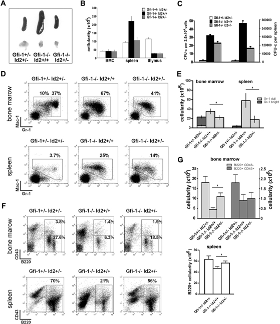
Figure 6.
Id2 heterozygosity partially rescues Gfi-1−/− phenotype. (A) Picture of spleens and thymuses from Gfi-1+/−Id2+/−, Gfi-1−/−Id2+/+, and Gfi-1−/−Id2+/− mice. Image taken with Canon PowerShot G10 Digital Camera. (B) Cellularity of bone marrow, spleens and thymuses from Gfi-1+/−Id2+/−, Gfi-1−/−Id2+/+, and Gfi-1−/−Id2+/− mice. (C) Splenocytes from Gfi-1+/−Id2+/−, Gfi-1−/−Id2+/+, and Gfi-1−/−Id2+/− mice were plated in methylcellulose medium supplemented with murine IL-3 and mGM-CSF. Colony numbers were counted after 7 to 10 days in culture. (D-E) Bone marrows and spleens of Gfi-1+/−Id2+/−, Gfi-1−/−Id2+/+, and Gfi-1−/−Id2+/− mice were analyzed for Gr-1 and Mac-1 expression by FACS. The percentage and total cell number of myeloid populations were shown. *P < .05 in 2-sided t test. (F-G) Bone marrows and spleens of Gfi-1+/−Id2+/−, Gfi-1−/−Id2+/+, and Gfi-1−/−Id2+/− mice were analyzed for B-cell development by FACS. The percentage and total cell number of B220+CD43+, B220+CD43−, or B220+ cells were shown. *P < .05 in 2-sided t test. Three independent experiments were performed.

