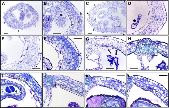
Figure 2.
Changes in the structure of pistil and fruit during their post-anthesis development. A to H, Transversal sections of cer6-2 pistil; anthesis (A and B) showing all cellular layers; 4 DPA (C) showing increased size and disorganization of the transmitting tract; 6 DPA (D) with degraded ovules (asterisk); 8 and 9 DPA (E and F); 10 DPA (G) showing septum degradation, sclerenchyma in the adaxial subepidermal layer (thick arrow), and adaxial epidermis collapse (thin arrow); and 11 DPA (H) showing lignification in the dehiscence zone (squared areas). I to L, Ovary transversal sections of cer6-2 seeded fruits; 8 DPA (I) with sclerenchyma in the endocarp a (squared area); 9 DPA (J) with endocarp b fully collapsed (squared area) and dehiscence zone formed (arrow); and 10 and 11 DPA (K and L). b, Abaxial epidermis; c, chlorenchyma; d, adaxial epidermis; e, seed; o, ovule; r, replum; s, septum; t, transmitting tract; arrowhead, stomata. Scale bar is 50 μm.

