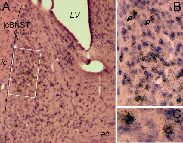
Figure 6.
Double in situ hybridization for GAD65 and GAD67 and for CRF in the dorsal BNST. A, A large number of GAD-containing neurons (shown in purple) are present in the jcBNST and in the dorsolateral BNST in general. In situ hybridization signal for CRF (brown grains) was seen in the dorsolateral BNST, including the jcBNST, and were primarily colocalized with GAD65 and GAD67. LV, Lateral ventricle. B, Higher magnification of the area in the dotted box in A shows high level of colocalization of CRF with GAD65 and GAD67 in the midsection of the jcBNST. C, Higher magnification of the two neurons containing CRF and GAD65 and GAD67 signal marked by the arrows in B.

