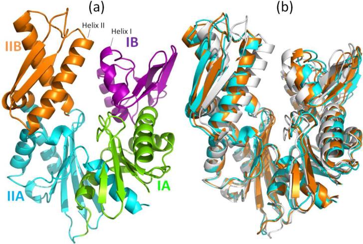
Figure 1.
(a) Subdomains of Hsp70 ATPase domain: IA (green; residues 1-39 and 116-188), IB (purple; residues 40-115), IIA (cyan; residues 189-228 and 307-C-terminus) and IIB (orange; residues 229-306). (b) Ribbon diagram of the superimposed closed and open conformations. The closed form (white) is a structure observed in the absence of nucleotide (PDB id: 1hpm) [16]. Two open forms are shown, both observed in the complexes formed with NEFs: In cyan is the structure from the complex with the Sse1 (PDB id: 3c7n) [17]; and in orange is that assumed when complexed with BAG (PDB id: 1hx1) [9]. The NEFs (Sse1 and BAG) are not shown here. The three structures have been aligned using the Kabsch algorithm ([21] as implemented in PyMol).

