
Figure 1.
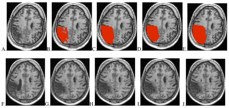
Methods of lesion masking in approximately aligned axial slices from one MCA stroke patient. Upper images (A-E): lesion in native space and shown with four masking conditions. Lower images (F-J): results after spatial normalization.
A. Native space lesion without mask B. Native space brain with unsmoothed, precise lesion mask C. Native space brain with smoothed, precise lesion mask D. Native space brain with unsmoothed, rough lesion mask E. Native space brain with smoothed, rough lesion mask F. Normalized brain using no lesion mask G. Normalized brain using unsmoothed, precise lesion mask H. Normalized brain using smoothed, precise lesion mask I. Normalized brain using unsmoothed, rough lesion mask J. Normalized brain using smoothed, rough lesion mask