Figure 3.
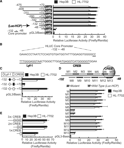
CREB-binding site contributes to the activation of the promoter. (A) Hep3B and HL-7702 cells were transiently co-transfected with reporter plasmids containing truncated versions of the promoter region of the Hulc gene, as indicated, and pRL-TK. Luc-HCP is defined as the reporter containing the shortest promoter region (from −132 to +72), which core elements located in. (B) Transcription factor binding sites present in the core promoter region (from −132 to −48) were predicated by web software TFSEARCH and MatInspector, as indicated by dashed lines. (C) Cells were transiently transfected with wild-type Luc-HCP as well as Luc-HCP which Lyf-1 or CREB binding site was deleted. Lyf-1-binding site is indicated by circle, whereas CREB-binding site is indicated by rectangle; deleted Lyf-1 and CREB sites are shown as black cycle and rectangle, respectively. (D) Scanning mutational analysis of the fragment from −132 to 48. As shown on the upper panel, a series of 8-bp substitutions were made within the fragment from −132 to 48 of the reporter construct Luc-HCP (from −132 to +72). M6, M7 and M13 overlap the possible CREB-binding site. (E) Wild-type Luc-HCP and Luc-HCP containing additional one (2×) or two (3×) possible CREB-binding sites were co-transfected with pRL-TK into Hep3B and HL-7702 cells. Firefly luciferase activity was normalized to Renilla luciferase activity, and the relative luciferase activities are presented as fold increase over the promoter-less pGL3 basic vector. Horizontal column lengths represent the means ± SD of three independent experiments.

