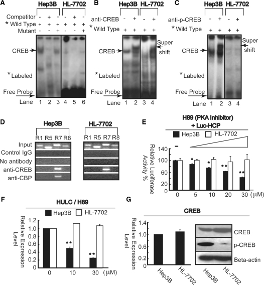
Figure 4.
HULC expression is regulated by phospho-CREB through PKA pathway in liver cancer cells. (A) Upon interaction with the nuclear extract of Hep3B (left panel) and HL-7702 (right panel), wild probe generated specific band (lanes 1 and 4) which was self-competed by 30-fold excess of unlabeled probe (lanes 2 and 5). No competition was observed when using the same amounts of mutant probe (lanes 3 and 6). (B) Interaction with anti-CREB antibody resulted in a super shift band formation in both two cell lines (lanes 2 and 4). (C) Interaction with anti-phospho-CREB antibody resulted in a super shift band formation (lane 2) in Hep3B, but not detected in HL-7702 cell line (lane 4). (D) ChIP analysis was performed to qualitative confirm the interaction of CREB and CBP with the Hulc promoter in vivo in Hep3B (upper panel) and HL-7702 (lower panel) cells using primer sets R1, R5, R7 and R8 described in Figure 2B. PCR products from the ChIP assay were run on an agarose gel. As the negative controls, the protein–DNA complexes were incubated without antibodies or with non-specific control IgG. The input DNA represents one-fifth of the starting material. (E) Pre-incubation with H89 was performed 1 day before transiently co-transfected Luc-HCP and internal control pRL-TK plasmids into Hep3B and HL-7702 cells and further incubation for 1 day in the continued presence of indicated amount of H89. Firefly luciferase activity was normalized to Renilla luciferase activity. Results are shown as relative percentage to those of cells untreated H89. (F) Total RNA was extracted for measurement of HULC mRNA expression level after treatment by indicated amount of H89 by real-time PCR. *P <0.05; **P <0.01 versus the corresponding untreated cells (E and F). (G) Endogenous CREB mRNA was quantified by real-time PCR and normalized to β-actin RNA in Hep3B and HL-7702 cells (left panel). The whole lysates of Hep3B and HL-7702 cells were examined by immunoblotting with antibodies against CREB, p-CREB and β-actin (right panel).

