Figure 3.
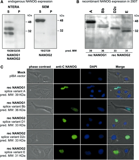
Western blot experiments, recombinant expression of NANOG1 and NANOG2 variants in 293T cells and immunohistological experiments. (A) Western blot experiments using an antiserum against the C-terminal portion of NANOG protein revealed the expression of NANOG1 and NANOG2 protein variants in NTERA-2 cells, and NANOG2 in SEM cells. (B) Cloned Strep-affinity-tagged splice variants of NANOG1 and NANOG2. Predicted molecular weight of all expression constructs is indicated. Recombinant expression of NANOG1 splice variants A and Bb and NANOG2 splice variants D2c and E in 293T cells were shown. Sizes of protein markers are indicated. (C) Immunohistological analysis of all recombinant NANOG1/2 protein variants expressed in Hela cells. Selected pictures from transfected cells were taken (20×). From left to right: phase contrast, anti-C NANOG antiserum, DAPI counter-staining and a merged picture is shown. All NANOG1/2 protein variants were localized within the nucleus of transfected cells.

