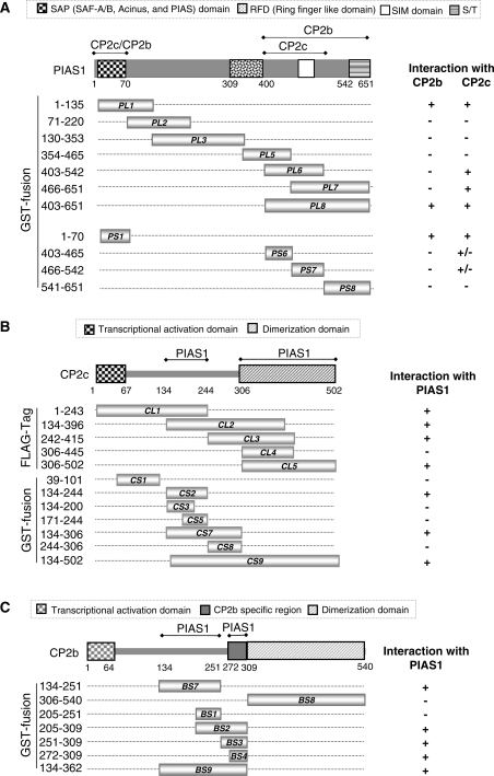
Figure 6.
Determination of binding regions joining PIAS1 and CP2c/CP2b. (A) Schematic representation of the PIAS1 deletion mutants and their binding capacities for CP2c and CP2b. The amino acids corresponding to the truncated PIAS1 proteins are indicated. Binding ability of each deletion mutant for CP2b or CP2c is indicated by plus (+) and minus (−) signs. Whole cell extracts from 293T cells expressing the HA-CP2c or EGFP-CP2b protein were mixed with each of the purified GST-tagged PIAS1 deletion mutants or GST and then subjected to pull-down analysis (Supplementary Figure S2A). PL and PS indicate the large and small fragments of PIAS1, respectively. (B) Schematic representation of CP2c deletion mutants and their binding capacities for PIAS1. Total cell extracts from 293T cells expressing the large fragments of FLAG-CP2c (CL1–CL5) or FLAG-PIAS1 were incubated with GST-PIAS1 or GST-CP2c deletion mutants (CS1–CS9), as indicated. GST pull-down proteins were analyzed by immunoblotting against anti-FLAG or GST antibodies (Supplementary Figure S2B). (C) Schematic representation of CP2b deletion mutants and their binding capacities for PIAS1. Whole cell extracts from 293T cells expressing FLAG-PIAS1 were mixed with purified GST-tagged small fragments of CP2b (BS) or GST, as indicated, and binding activities were analyzed in GST pull-down assays, followed by immunoblotting with FLAG or GST antibodies (Supplementary Figure S2C).

