Figure 4.
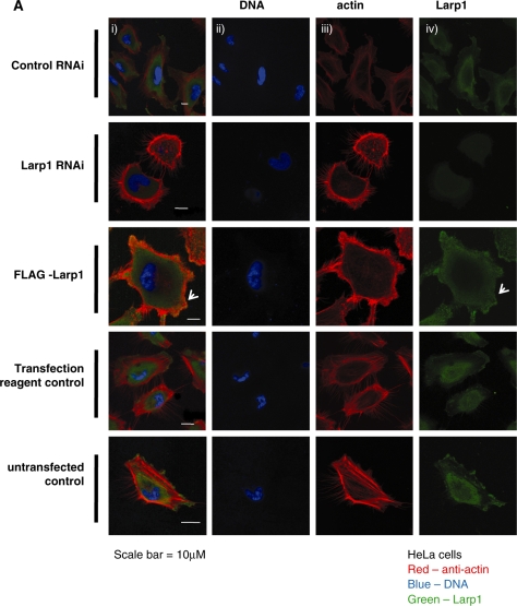
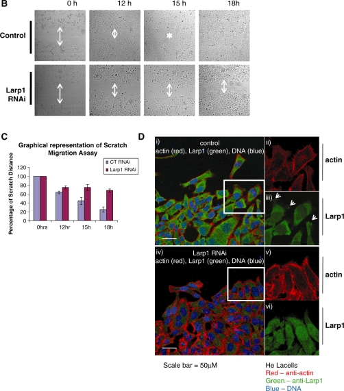
(A) Immunofluorescence of HeLa cells following treatment with control RNAi, Larp1 RNAi and in cells transfected with Larp1 over-expressing construct (FLAG-Larp1) examined using confocal microscopy. Panels show images in combined colour channel (i), blue channel (DNA) (ii), red channel (actin) (iii) and green channel (Larp1) (iv). Larp1 over-expression (FLAG-Larp1) causes a mesenchymal-like phenotype, with cells displaying multiple lamellipodia (white arrow). Cells treated with Larp1 RNAi were larger and had more filopodia when compared to controls. (B) Scratch migration assay in HeLa cells treated with Larp1 siRNA or control siRNA control. An open furrow was generated by scratching confluent cells using a pipette tip. Confluency was restored in controls between 15 and 18 h. However, in cells treated with Larp1 siRNA, confluency was not restored after 18 h. The distance between furrow edges in the control and Larp1 siRNA treated cells was measured and is presented graphically in (C). In (D), cells were fixed and immunostained 12 h after scratch. Cells at the furrow edge were examined using confocal microscopy. This showed, in cells treated with control RNAi (top panel), a concentration of Larp1 within their leading edge (small arrows in magnified figures) in the direction of cell travel. This was not observed after Larp1 RNAi (bottom panel), where cells were closely adherent to each other without a clear direction of travel.


