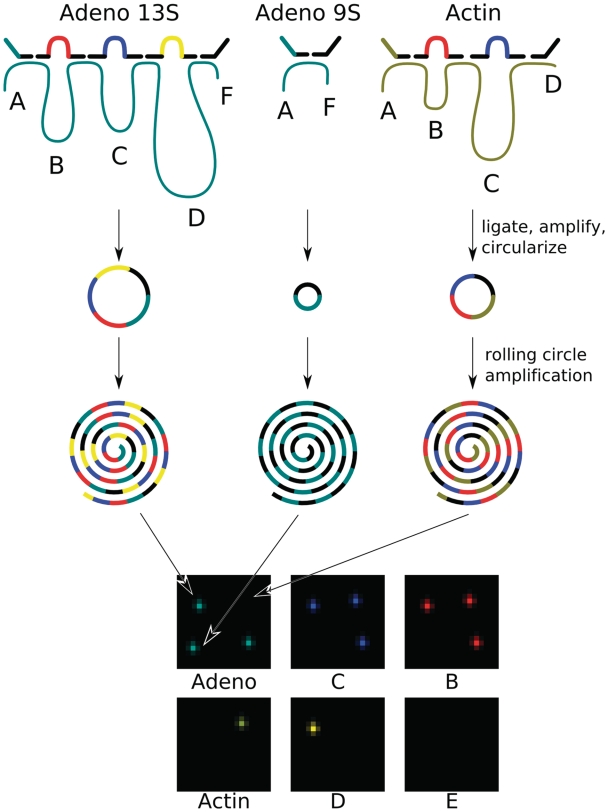
Figure 2.
The spliceotyping detection scheme: exon-specific probes hybridize to the adenovirus (dark cyan) and actin (brown) cDNAs and are ligated into circular DNA tag strings that are amplified and circularized. Each tag string derived from an individual transcript is amplified by RCA, encoding the exon content of the transcript. RCPs are spread out on a glass slide and decoded by sequential hybridization with probes directed against the tag sequences. The pictures show combined data from the three decoding steps where the adenovirus and actin gene-id tags are decoded together with the exon tags B, C, D and E.

