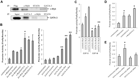
Figure 3.
GATA-3 and c-Myb act cooperatively to activate the GATA-3 promoter. (A) Western Blots of immunoprecipitation (IP) carried out on lysates from human primary CD4+CD45RO− cells stimulated for 3 to 5 days under Th2 cell-promoting conditions. Flag indicates Flag-tagged c-Myb expressed in Jurkat cell lysate and IP with anti-Flag antibody (positive control); and Input, total GATA-3 present in cell lysate from stimulated CD4+ T cells. The blots shown are representative of 3 independent experiments. (B) Reporter assays carried out in 293T cells 48 hours after transfection with pG3P-S (wild-type Myb binding site) in the presence of c-Myb and/or GATA-3 expression constructs, or control (empty) vectors. Data in the left and right graphs are representative of 2 and 5 independent experiments, respectively. (C) Reporter assays carried out with pG3P-S or pG3P-M (mutated Myb binging site) as described in Figure 2C. (D) Reporter assays performed in human peripheral T cells with pG3P-S in the presence of c-Myb and/or GATA-3 expression constructs, or (E) siRNA against c-myb, 24 hours after transfection. (C-E) Data are representative of at least 2 independent determinations. *P < .05. **P < .01.

