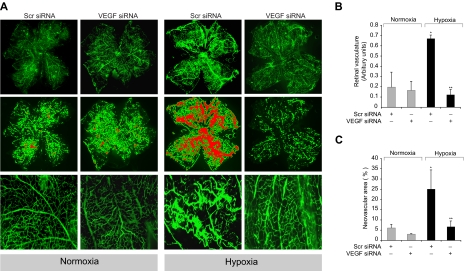
Figure 8.
Depletion of VEGF levels reduces hypoxia-induced retinal neovascularization. After exposure to 75% oxygen, the mice pups were returned to normoxia and administered 1 μg of Scr or VEGF siRNA at P13, P14, and P15 by intravitreal injections. Pups were anesthetized at P17, perfused with FITC-dextran, sacrificed, enucleated, retinas were isolated, and flat mounts of whole retina were examined for retinal neovascularization (A). Neovascular tufts were highlighted with red (second row). The third row shows the magnified (original magnification ×10) section of a selected area of the images shown in the first row (original magnification ×2). The images were captured using an Inverted Zeiss fluorescence microscope (AxioVision AX10), and fluorescence intensity in the total retinal area (B) and the area of neovascular tufts to total retinal area (C) was analyzed by Nikon NIS-Elements software Version AR3.1. Values are mean ± SD. *P < .01 versus normoxia plus scrambled siRNA control. **P < .01 versus hypoxia plus scrambled siRNA.

