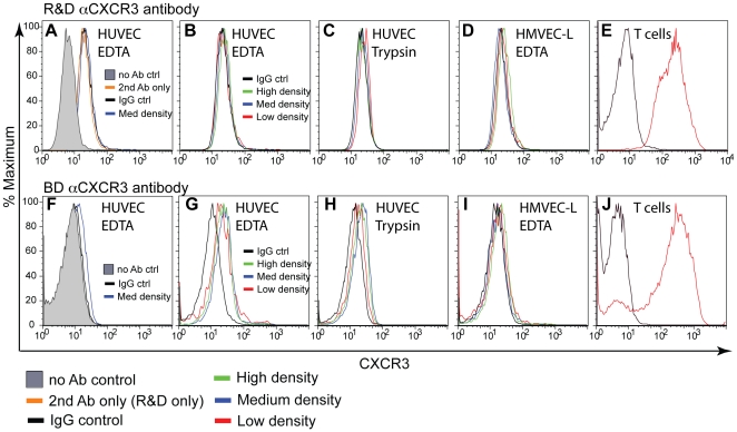
Figure 5. HUVEC and HMVEC-L have no detectable surface CXCR3 expression.
CXCR3 surface levels were assessed by flow cytometry using anti-hCXCR3 monoclonal antibodies from R&D (A–E), or BD Bioscience (F–J), for HUVECS (A–C, F–H), HMVEC-L (D and I) and human peripheral blood T cells cultured in IL-2 (E and J). Endothelial cells were harvested at different densities to determine cell cycle dependency of CXCR3 expression (B–C, G–H), and were harvested either with EDTA (A, B, D, F, G, I) or trypsin (C and H).

