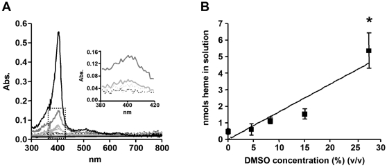
Figure 1. DMSO promotes spontaneous heme solubilization in acidic conditions.
(A) Different concentrations of DMSO in 0,5 M sodium acetate buffer pH 4.8 and 100 µM heme with a final volume of 1.0 mL were shaken for 10 minutes and centrifuged at 10 000×g. for 10 min. The supernatants were analyzed by uv-visible spectroscopy between 300 nm and 800 nm. An expansion magnification of the dotted box is shown in the inset. Dashed line black: control; dashed line gray: 4.6% DMSO; pale gray: 8.3% DMSO; dark gray: 15.1% DMSO; black: 27.7% DMSO. (B) Heme content in solution was quantified using the alkaline pyridine method. Data are expressed as mean ± SEM, of three different experiments in B.

