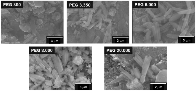
Figure 5. Scanning electron micrographs of βH induced by PEGs.
Scanning electron microscopy (SEM) was used to investigate the external morphology of the βH crystals produced by different PEGs. Well formed crystals are seen in the presence of PEG 6.000, 8.000 and 20.000 which closely resemble hemozoin. Less regular crystals appear to be formed by PEG 3.350 and few if any are formed in the presence of PEG 300.

