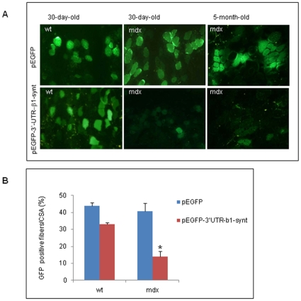
Figure 4. Gene delivery into tibialis muscles of wt and mdx mice.
A: Micrographs of GFP expression in the muscle sections from tibialis muscles of wt or mdx mice injected with 20 µg of DNA containing either pEGPF-C1 or pEGFP-3′-UTR-β1-synt, and subjected to electric pulses. A representative experiment is shown. B: The graph values represent the mean ± SE of the percentage of GFP positive cells/muscle sections analyzed in 30-day old wt and mdx mice. Fifteen sections per experimental group (n = 3 wt, n = 4 mdx) were counted. *P<0,05 as measured by the unpaired t-test.

