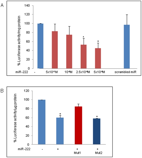
Figure 6. Luciferase activity in the presence of miR-222.
A: Luciferase activity in COS1 cells transfected with pGL3-3′-UTR-β1-syntrophin was assessed in the absence or presence of different concentrations of miR-222 and or antimiR (5×10⁻⁸M). Luciferase activity was normalized to total protein levels and calculated as the percentage of the values obtained from miR-transfected samples compared to those obtained from untreated cells. Results are presented as the mean ± SE from five independent experiments. Statistical significance was determined as *P<0,05 versus untreated cells (P = 0,0045 [2.5×10⁻⁸M], P = 0,0027 [5×10⁻⁸M] measured by the Mann-Whitney test). B: Luciferase activity of COS1 cells was assessed in cells transfected with pGL3-3′UTR-β1-syntrophin or two vectors with mutations in the first (Mut1) or second (Mut2) putative binding site for miR-222 in the absence or presence of 5×10⁻⁸M of miR-222. Results are presented as the mean ± SD from three independent experiments. *P<0,05 versus untreated cells (P = 0,040 measured by the Mann-Whitney test).

