Figure 2.
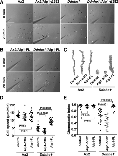
Impaired chemotaxis of Ddnhe1− cells is enhanced by DdAip1-Δ382 but suppressed by DdAip1-FL. Chemotactic movement toward a micropipette tip filled with cAMP was recorded for 30 min. (A) Images of Ax2 and Ddnhe1− cells without and with heterologous expression of DdAip1-Δ382. (B) Images of Ax2 and Ddnhe1− cells expressing DdAip1-FL. (C) Morphology and tracking of the indicated cell types determined by drawing and overlapping images of a single cell from frames taken at 1-min intervals. (D) Speed of the indicated cell types, expressed as total distance moved divided by total moving time. The speed of Ddnhe1− cells is significantly decreased by DdAip1-Δ382 but increased by DdAip1-FL. (E) chemotactic index of the indicated cells types, determined as net distance moved divided by total moving distance during the time period. The chemotactic index of Ddnhe1− cells is significantly decreased by DdAip1-Δ382 but increased by DdAip1-FL. Each dot in the scatter plots represents an individual cell in time-lapse images, and data are representative of three independent experiments.

