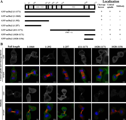
Figure 2.
Analysis of the localization of mDia2 deletion mutants during cell division. (A) A schematic representation of the constructs and summary of the localization analysis. Experiments were carried out as described in B. More than 20 cells from two or three experiments were analyzed to evaluate the localization at the cleavage furrow, central spindle, and midbody. FH1 or FH2, formin homology domain 1 or 2, respectively; DID, diaphanous inhibitory domain; DAD, diaphanous auto-regulatory domain, GBD, GTPase-binding domain; ARR, Armadillo repeat domain; DD, dimerization domain. X indicates the position of mutation. I704A, the actin-polymerization defective mutation (Watanabe et al., 2008). (B) Localization of each GFP-mDia2 deletion mutants during cell division in NIH 3T3 cells. NIH 3T3 cells were transfected with a series of GFP-mDia2 deletion mutants, and the cells were fixed and stained for GFP (green), microtubules (red), and DNA (blue). Bar, 5 μm.

