Figure 5.
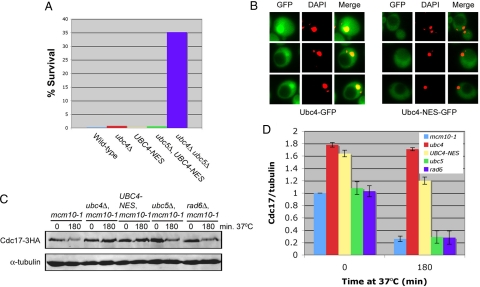
Cdc17 is degraded primarily in the nucleus. (A) ABy150 (S288C), ABy152 (ubc4Δ), ABy574 (UBC4-NES-3HA), ABy577 (ubc5Δ, UBC4-NES-3HA), and GAP510 (ubc4Δ, ubc5Δ) were heat shocked as described in the Materials and Methods, aliquots were grown on YPD plates for 3 d at 30°C, and colonies were counted to determine the percentage of cells that were viable following heat shock. (B) ABy605 (mcm10-1, UBC4-GFP) and ABy660 (mcm10-1, UBC4-NES-GFP) were examined using fluorescence microscopy. Merge represents GFP and DAPI. Yellow dots represent colocalization of Ubc4-GFP and DAPI-stained nuclei. Red dots represent DAPI-stained nuclei in which Ubc4-GFP is not present. Black circles represent vacuoles. (C) Asynchronous cultures of ABy013 (mcm10-1), ABy342 (mcm10-1, ubc4Δ), ABy455 (mcm10-1, UBC4-NES-3HA), ABy363 (mcm10-1, ubc5Δ), and ABy359 (mcm10-1, rad6Δ) grown at 25°C were shifted to 37°C for 180 min. Cdc17–3HA and α-tubulin were analyzed by Western blot. (D) The graph shows Cdc17/tubulin ratios at each time point for each strain relative to mcm10-1 at time 0 (average of 3 separate experiments, bars represent mean ± SD).

