
Figure 5.
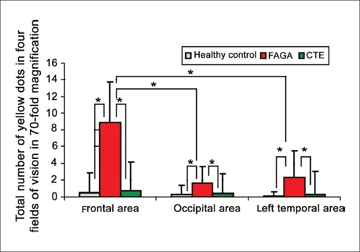
Yellow dots in frontal, occipital and left temporal areas of all three groups of patients, presented as a number counted in four field of visions at 70-fold magnifications. Asterix marks statistically significant differences (P < 0.001)
Official websites use .gov
A
.gov website belongs to an official
government organization in the United States.
Secure .gov websites use HTTPS
A lock (
) or https:// means you've safely
connected to the .gov website. Share sensitive
information only on official, secure websites.
Yellow dots in frontal, occipital and left temporal areas of all three groups of patients, presented as a number counted in four field of visions at 70-fold magnifications. Asterix marks statistically significant differences (P < 0.001)