Figure 2.
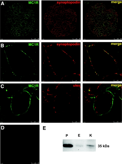
MC1R is expressed in normal human kidney tissue. MC1R colocalized with synaptopodin in a podocyte-specific manner. (A) Confocal microscopic analysis of cryosections from human kidney tissue confirmed expression of MC1R (green) and synaptopodin (red). Colocalization of these proteins was shown in merge (yellow). (B) Enlarged images of one glomerular capillary loop. (C) MC1R (green) and the endothelial-specific lectin UEA I (red) did not colocalize (merge). (D) Omission of primary antibody yielded no staining. Scale bar = 50 μm. (E) Western blotting was performed with an anti-MC1R antibody. MC1R was detected in high amounts primarily in podocytes. P, podocytes; E, endothelial cells; K, kidney tissue.

