Figure 2.
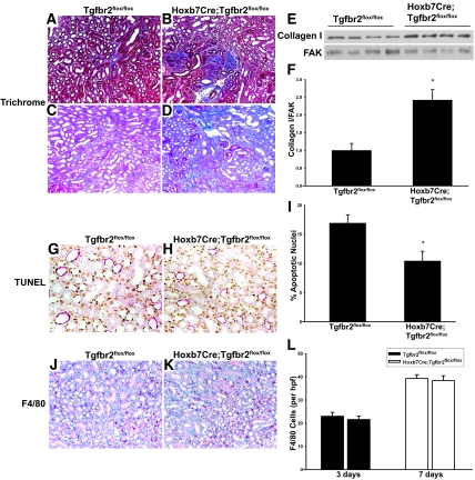
Hoxb7Cre;Tgfbr2flox/flox mice have increased collagen I expression that is not attributed to changes in apoptosis or inflammation. (A through D) Fibrillar collagen is detected 7 (A and B) and 14 (C and D) days after UUO by Trichrome staining. (E) Lysates of corticomedullary tissue of obstructed kidney are immunoblotted for collagen I. (F) Collagen I bands from four mice per genotype are quantified by densitometry, normalized to focal adhesion kinase (FAK), and reported as means ± SEM. *P < 0.05. (G and H) TUNEL staining shows apoptotic nuclei (brown) in Hoxb7Cre;Tgfbr2flox/flox and Tgfbr2flox/flox mice 3 days after UUO. CD localization of apoptosis is determined by co-staining with AQP2 (red). (I) TUNEL-positive cells from 10 high-powered fields per mouse are counted in a blinded manner using four mice per genotype. *P < 0.05. (J and K) F4/80 staining is performed for macrophage infiltration. (L) F4/80-positive cells are quantified and expressed as described for TUNEL staining. Magnifications: ×100 in A through D, J, and K; ×400 in G and H.

