Figure 4.
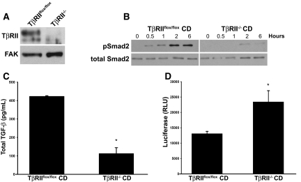
Conditioned media from TβRII−/− CD cells have greater levels of active TGF-β than TβRIIflox/flox CD cells. (A) TβRII−/− and TβRIIflox/flox CD cell lysates are immunoblotted with a TβRII antibody. (B) TβRIIflox/flox and TβRII−/− CD cells are stimulated with TGF-β (5 ng/ml) for various periods, and then immunoblots for pSmad2 are performed. (C and D) Total and active levels of TGF-β from media of TβRIIflox/flox and TβRII−/− CD cells collected over 72 hours are determined by ELISA (C) and PAI/L assays (D). Total TGF-β (C) is reported as pg of TGF-β per mg of protein, and active TGF-β (D) is measured by random luciferase units (RLU). The values reported in C and D reflect averages ± SE from three separate experiments. *P < 0.05.

