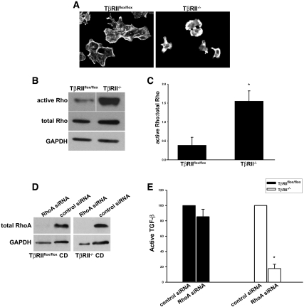
Figure 7.
TβRII−/− CD cells have increased active RhoA. (A) Rhodamine phalloidin staining of actin cytoskeleton in TβRIIflox/flox CD and TβRII−/− CD cells. (B) Rho activity in cell lysates is measured as described in the Concise Methods section, and a representative experiment (a total of three performed) is shown. (C) A ratio of the densitometry of active Rho to total Rho from these three experiments is expressed as mean ± SEM. *P < 0.05. (D) Lysates of CD cells transfected with either pooled RhoA siRNA or control siRNA are immunoblotted with an antibody to RhoA. (E) Active TGF-β in the conditioned medium of cells transfected with RhoA siRNA is measured using the PAI/L assay and expressed as a percentage of active TGF-β measured in control siRNA-treated CD cells. This experiment was performed three times with error bars depicting the SE. *A significant (P < 0.05) drop in active TGF-β in RhoA siRNA-treated TβRII−/− compared with TβRIIflox/flox CD cells.

