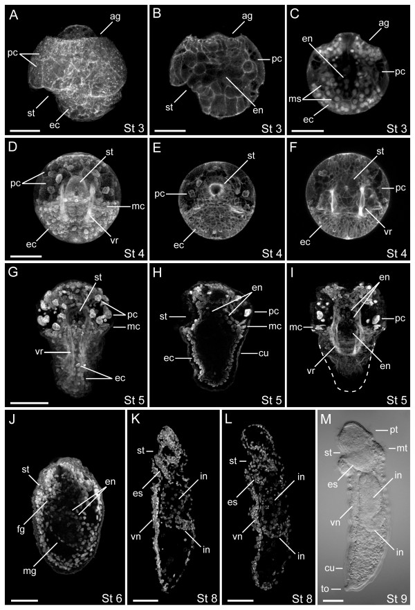
Figure 3.
Micrographic analysis of gut formation in the sipunculan, Themiste lageniformis. (A-L) confocal laser scanning micrographs (CLSM). Actin filaments are labeled with BODIPY phallacidin (A-I); cell nuclei are labeled with anti-histone antibody (C-J) or propidium iodide (K, L); (M), light micrograph with DIC optics. (A, B, H, J-M) lateral views with ventral to the left and anterior up; (C, I) dorsal views with anterior up; (D-G) ventral views with anterior up. (A) Three dimensional z-series of a stage 3 embryo showing position of the stomodeum. (B) Single focal plane of the embryo in (A). (C) Dorsal view z-series of a stage 3 embryo showing relative positions of ectoderm, mesoderm and endoderm cells. (D) Three dimensional z-series of a stage 4 embryo before elongation. The stomodeum is closed at its dorsal end. (E, F) Two z-series composites of the same embryo in (D), at progressively deeper focal planes. The stomodeum forms a 'bowl' that is lined with ciliated cells, and terminates with a rosette of larger cells. (G-I) Ventral, lateral and dorsal views of three different conical-shaped stage 5 embryos. The stomodeum is oriented parallel to the D/V axis just anterior to the metatroch. (J) Stage 6 larva with the stomodeum in an anterior to posterior orientation, and midgut with endoderm nuclei. (K, L) Early and late stage 8 pelagosphera, respectively. The esophagus is packed with cells and the intestine ascends toward the dorsal body wall. (M) Stage 9 pelagosphera after 4 days of growth. Descending and ascending arms of the intestinal system are now visible. ag, apical groove; cu, cuticle; ec, ectoderm cells; en, endoderm cells; fg, foregut; hg, hindgut; mc, metatroch cells; mg, midgut; ms, mesoderm cells; mt, metatroch; pc, prototroch cells; pt, prototroch; st, stomodeum; to, terminal organ; vn, ventral nerve cord; vr, ventral retractor muscle. Scale bars = 50 μm.

