Figure 5.
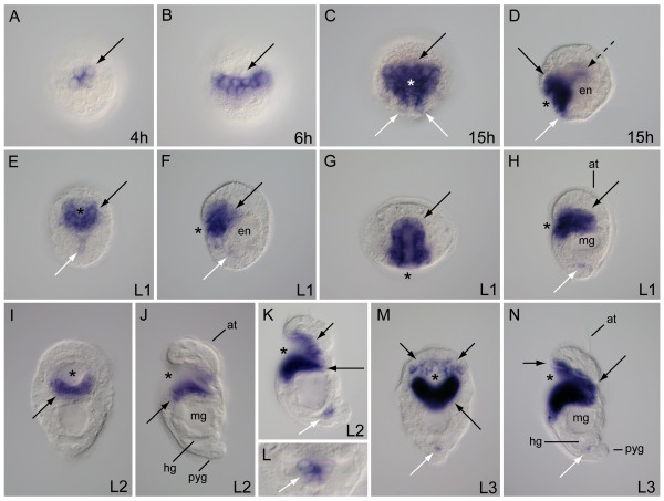
Developmental expression of FoxA in Chaetopterus. (A, B) Vegetal views; (C, E, I, M) ventral views with anterior to the top (D, F, H, J, K, N) lateral views with anterior to the top and ventral left; (G) anterior view with ventral down. (A) FoxA is expressed in a group of four vegetal cells (arrow) 4 hours after fertilization. (B) Late gastrula-stage embryo 6 hours after fertilization with expression (arrow) on the vegetal plate. (C) Protrochophore larva at 15 hours showing FoxA expression in the stomodeum (black arrow) and a pair of ventral posterior cells (white arrows). (D) Same larva as in C, showing FoxA expression in the stomodeum (solid black arrow), posterior to the stomodeum (white arrow) and in endoderm (broken black arrow). (E, F) L1 larva at 25 hours with FoxA transcription in the stomodeum (black arrow) and subsurface cells (white arrow) along the ventral midline. (G) L1 at 27 hours with FoxA expression (black arrow) lining the stomodeal canal. (H) Same larva as in (G), showing expression in the stomodeum (black arrow) and putative hindgut cells (white arrow). (I, J) L2 larva showing positive FoxA cells (black arrow) in the posterior side of the stomodeum. (K) Extended color development in L2 larva with expression along the ventral posterior side of the stomodeum (long black arrow), within the stomodeum roof (short black arrow) and in the rectum (white arrow). (L) Posterior dorsal view of larva in K. FoxA-positive cells (white arrow) surround the rectal canal. (M, N) L3 larva showing FoxA expression along the stomodeum canal (long black arrow), in the stomodeum roof (short black arrows) and the rectal canal (white arrow). Asterisk marks position of the stomodeum. h, hours post fertilization; at, apical tuft; en, endoderm; hg, hindgut; mg, midgut; pyg, pygidium.

