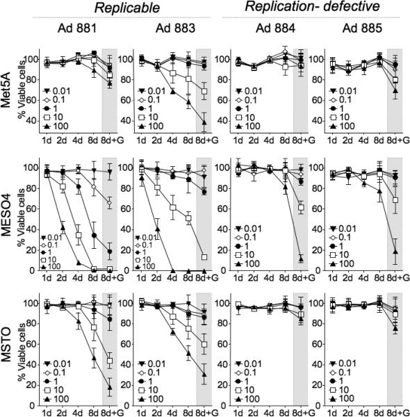
Figure 6. Quantitative in vitro cytotoxicity assay.
Met5A, MESO4, and MSTO cells (1 × 10⁴/well) were cultured in multiple replicate 96-well plates and infected with Ad881, Ad883, Ad884, or Ad885, at MOIs from 0.01 to 100. On the indicated days, the number of surviving cells was analyzed by a colorimetric methods using Alamar blue. One set of cultures was treated with GCV (1 mg/mL) during the last three days prior to the colorimetric assay (8d+G). Data shown are the mean ± SD calculated from triplicates.

