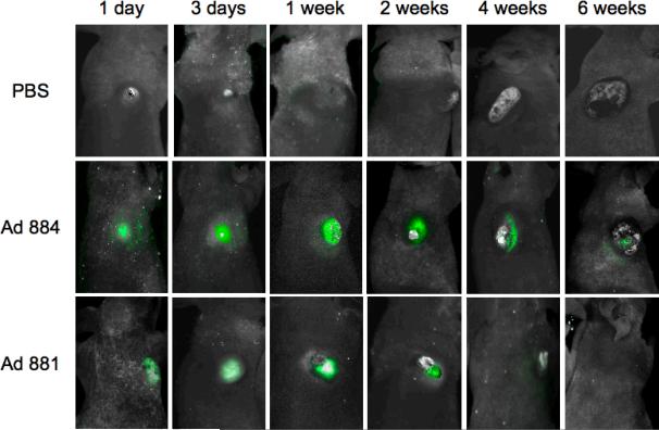
Figure 7. In vivo imaging of the spread of oncolytic adenovirus in malignant mesothelioma xenograft tumors.
MESO4 tumors were grown subcutaneously in nude mice to approximately 5–6 mm in diameter, and injected intratumorally with 5 × 10⁸ TU of either Ad881 or Ad884, or PBS control on Day 0 (n = 3 per group). At different time points indicated in the figure, whole body images (0.05- to 0.5-second exposure) were taken and analyzed by in vivo fluorescence imaging. Representative images are shown from each group.

