
Figure 7.
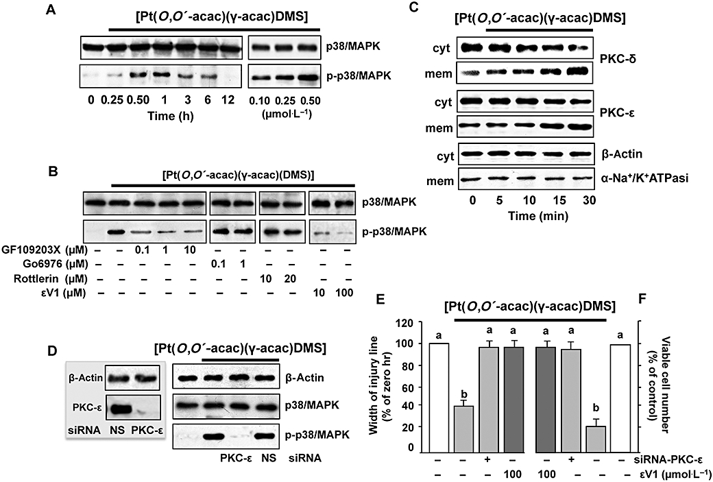
[Pt(O,O′-acac)(γ-acac)(DMS)] induces p38MAPK activation. (A) Cells pretreated or not with the PKC inhibitors, GF109203X, Gö6976, rottlerin or εV1, were treated or not with 0.50 µmol·L−1[Pt(O,O′-acac)(γ-acac)(DMS)] for 1 h. Cell lysates were analysed by Western blotting with anti-total-p38MAPK (unphosphorylated and phosphorylated p38MAPK) and phosphorylated p38MAPK antibodies. Control loading is shown by β-actin. Representative immunoblots of three experiments are depicted. (B) MCF-7 cells were treated without or with [Pt(O,O′-acac)(γ-acac)(DMS)] for the indicated times. Cell fractions (cytosol and membranes for translocation studies) were analysed by Western blotting with specific anti-PKC-ε and anti-PKC-δ antibodies. The purity of fractions was tested by immunoblotting with anti β-actin and anti-α subunit of Na+/K+ATPase monoclonal antibodies. The figures are representative of four independent experiments. (C,D) Cells were transfected with siRNA–PKC-ε or control siRNA (NS) and then were incubated with 0.50 µmol·L−1[Pt(O,O′-acac)(γ-acac)(DMS)]. (C) Western blotting of total lysates was performed with specific anti-PKC-ε (inset) or with anti-unphosphorylated (p38MAPK) and phosphorylated p38MAPK (p-p38MAPK) antibodies. Control loadings are shown by β-actin. Representative immunoblots of three experiments are depicted. (D) Differential cell migration rate was examined using wound closure assay. Data are means ± SD of four different experiments and are presented as per cent of wound closure for control cells. (E) Cells were plated in polyHEMA plates. The number of surviving cells was determined, after 24 h, by an MTT assay and is expressed as per cent change from the initial seeding (time 0). (E,F) Values with shared letters are not significantly different according to Bonferroni/Dunn post hoc tests.