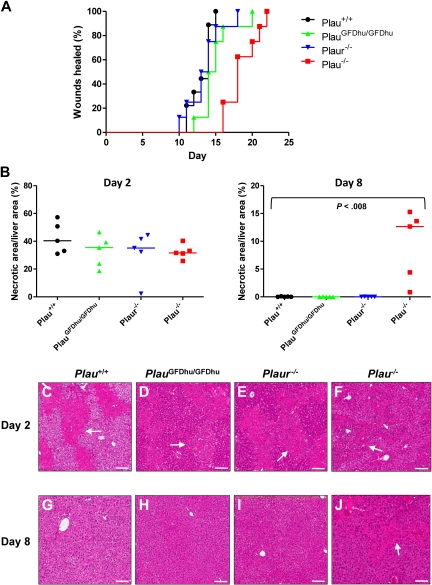
Figure 6.
Loss of uPA binding to uPAR does not affect tissue repair. (A) Rate of healing of 1.5-cm incisional skin wounds in Plau+/+ (black, N = 10), PlauGFDhu/GFDhu (green N = 10), Plaur−/− (blue, N = 10), and Plau−/− (red, N = 9) mice. Loss of uPA, but not uPA binding, to uPAR or loss of uPAR delays wound healing. P < .001 Plau−/− versus Plau+/+, PlauGFDhu/GFDhu or Plaur−/−, log-rank test, 2-tailed. (B) Area of hepatic necrosis 2 (left panel) and 8 (right panel) days after carbon tetrachloride–induced liver injury of Plau+/+ (black circles), PlauGFDhu/GFDhu (green triangles), Plaur−/− (blue triangles), and Plau−/− (red squares). A total of 5 mice were analyzed per genotype and time point. The extent of hepatic injury is similar in mice of all genotypes at day 2. At day 8, necrotic areas are completely cleared in Plau+/+, PlauGFDhu/GFDhu, and Plaur−/− livers, but not in Plau−/− livers. The P value was determined by the Mann-Whitney U test, 2-tailed. (C-J) Representative examples of the histologic appearance of livers from Plau+/+ (C,G), PlauGFDhu/GFDhu (D,H), Plaur−/− (E,I), and Plau−/− (F,J) mice 2 (C-F) and 8 (G-J) days after carbon tetrachloride administration. White arrows show examples of necrosis. Size bars, 100 μm.

