Figure 8.
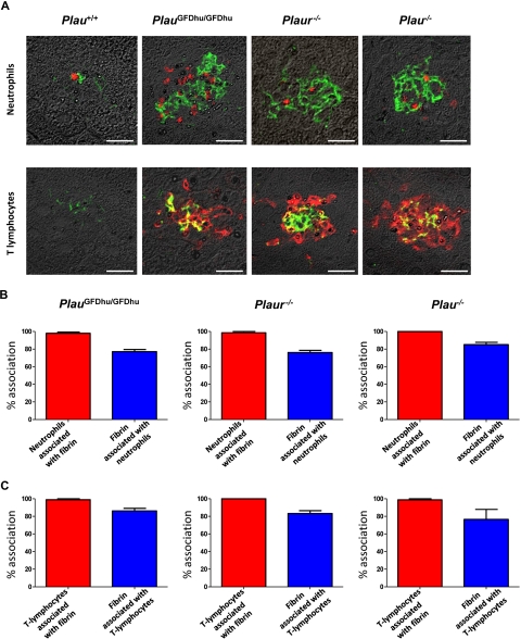
Inflammation caused by uPA-uPAR abrogation is secondary to fibrin deposition. (A) Examples of immunofluorescence using fibrin antibodies (green) combined with antibodies against neutrophils (myeloperoxidase, top panels) and T lymphocytes (CD3, bottom panels; red) of the livers from 1-year-old Plau+/+ (left panels), PlauGFDhu/GFDhu (2 left panels), Plaur−/− (2 right panels), and Plau−/− (right panels). Size bars, 20 μm. (B) Enumeration of the percentage of neutrophils associated with fibrin (red bars) and the percentage of fibrin deposits associated with neutrophils (blue bars) in the livers from PlauGFDhu/GFDhu (left), Plaur−/− (middle), and Plau−/− (right) panels. (C) Enumeration of the percentage of T lymphocytes associated with fibrin (red bars) and the percentage of fibrin deposits associated with T lymphocytes (blue bars) in the livers from PlauGFDhu/GFDhu (left), Plaur−/− (middle), and Plau−/− (right) panels.

