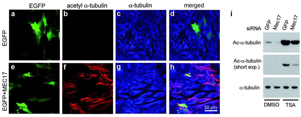
Figure 4. MEC-17 controls the levels of microtubule acetylation in mammalian cells.
a–h, Expression of Mm-MEC-17 in Ptk2 cells increases the levels of acetyl-K40 α-tubulin. Cells expressing either EGFP or EGFP and Mm-MEC17 were stained with 6–11 B-1 mAb and anti-α-tubulin antibodies. i, Depletion of Hs-MEC-17 in HeLa cells reduces the level of acetyl-K40 α-tubulin. Cells were transfected with either GFP or Hs-MEC17 siRNAs and after 50 hr, treated for 7 hr with either 300 nM trichostatin A (TSA, stock solution in DMSO) or DMSO alone. Cell lysates were analyzed by western blot probed with either 6–11 B-1 mAb (top, middle panels) or anti-α-tubulin mAb (bottom panel).

