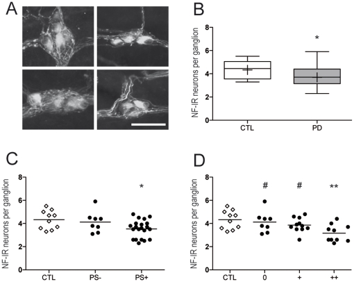
Figure 2. Count of neurofilament-positive neurons in the submucosal plexus of PD patients.
A. Neurofilament-immunoreactive (NF-IR) submucosal neurons were counted in every available ganglion from colonic biopsies. Representative photographs of ganglia from PD patients (left panels) and controls (right panels). Scale bar: 30 µm. B. A significant decrease in the number of NF-IR neurons per ganglion was present in the SMP of PD patients (PD, n = 29) as compared to controls (CTL, n = 10) (p<0.05). The bottom and the top of the box represent the 25th and 75th percentiles, respectively, and the end of the whiskers represent the minimum and maximum values; the median is represented as a bar and the mean as a ‘+’ sign inside the box. C. When segregating patients according to the presence (PS+) or absence (PS-) of phospho-synuclein IR neurites, the difference between patients and controls was sustained only for the group with Lewy pathology (PS+, n = 21, p<0.05). D. When further stratifying patients according to the density of pathology, the difference between patients and controls was sustained only for the group with severe Lewy pathology (++, n = 10, p<0.01). Groups without (0) or with moderate pathology (+) significantly differed from the group with severe (++) pathology (p<0.05). Each white square represents one control, each black circle represents one PD patient. Horizontal bars represent the mean. *p<0.05 and **p<0.01 as compared with controls. #p<0.05 as compared with the group with severe pathology (++).

