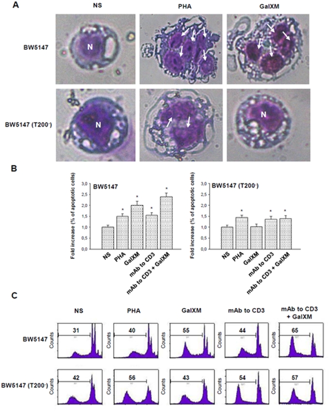
Figure 4. GalXM-induced apoptosis of BW5147 cells.
(A) BW5147 and BW5147 (T200−) cells (both 1×10⁶/ml) were incubated for 18 h in the presence or absence (NS) of PHA (10 µg/ml) or GalXM (10 µg/ml). After incubation, cells were collected by cytospin and stained by Hemacolor. GalXM-treated cells exhibited altered morphology, surface blubs and nuclear fragmentation (arrows). N = cell nucleus; original magnification 40x. In selected experiments, cells were incubated for 18 h in the presence or absence (NS) of PHA (10 µg/ml), mAb to CD3 (1 µg/ml) or GalXM (10 µg/ml). After incubation, the percentage of cells undergoing apoptosis was evaluated by PI staining and analyzed using FACScan flow cytometry. Data are expressed as fold increase of percentage of apoptotic cells (B), or shown as FACScan histograms from one representative experiment out of seven with similar results (C). *, p<0.05 (treated vs untreated, n = 7). Error bars denote s.e.m.

