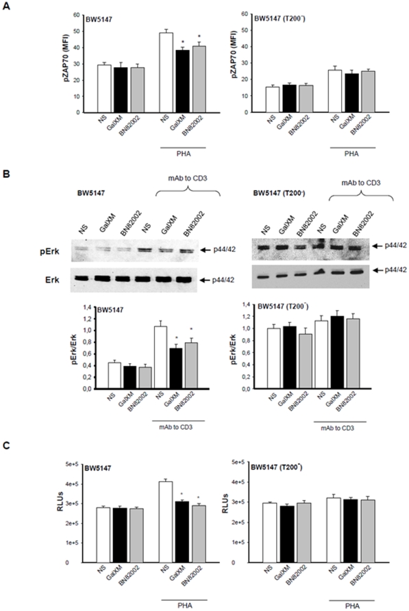
Figure 6. Phospho-ZAP70, phospho-Erk activation and proliferation of BW5147 cells treated with GalXM or BN82002.
(A) BW5147 and BW5147 (T200−) cells (both 1×10⁶/ml) were pre-activated for 30 min in the presence or absence (NS) of PHA (10 µg/ml) and then incubated for 30 min in the presence or absence (NS) of GalXM (10 µg/ml) or BN82002 (6 µM). After incubation cells were labelled with antibody to phospho-ZAP70 and then analyzed by FACScan flow cytometry. The mean of fluorescence intensity (MFI) of labelled cells is shown as a histogram. *, p<0.05 (treated vs untreated, n = 7). (B) BW5147 and BW5147 (T200−) cells (both 5×10⁶/ml) were pre-activated for 30 min in the presence or absence (NS) of mAb to CD3 (1 µg/ml) and then incubated for 30 min in the presence or absence (NS) of GalXM (10 µg/ml) or BN82002 (6 µM). After incubation, cell lysates were analyzed by Western blotting; membranes were incubated with antibodies to phospho-Erk1/2 and Erk1/2. pErk was normalized against Erk. *, p<0.05 (treated vs untreated, n = 7). Error bars denote s.e.m. (C) BW5147 and BW5147 (T200−) cells (both 1×10⁶/ml) were pre-activated for 30 min in the presence or absence (NS) of PHA (10 µg/ml) and then incubated for 48 h in the presence or absence (NS) of GalXM (10 µg/ml) or BN82002 (6 µM). After incubation, cell proliferation was evaluated by ViaLight Plus Kit. *, p<0.05 (treated vs untreated, n = 7). Error bars denote s.e.m.

