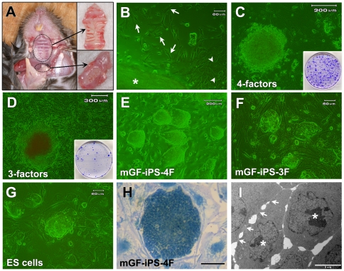
Figure 1. Generation of mouse GF-derived iPS cells.
(A) Palatal (upper inset) and mandibular (lower inset) gingival tissues of adult mice were extracted for establishment of primary GFs. (B) Fibroblasts (arrow) and epithelial cells (arrowhead) migrated out of the palatal gingival tissue (asterisk). Scale bar; 60 µm. (C) The morphology of a colony derived from mGFs 19 days after transduction of the four factors. Scale bar; 200 µm. Inset: Colonies in a 60-mm dish after staining with crystal violet (CV) on day 21. (D) The morphology of a colony derived from mGFs 49 days after transduction of the three factors. Scale bar; 300 µm. Inset: Colonies in a 100-mm dish after staining with CV on day 50. (E–G) Morphology of (E) mGF-iPS-4F-1 cells (Scale bar; 200 µm), (F) mGF-iPS-3F-1 cells (Scale bar; 60 µm) and (G) mouse ES cell line (Scale bar; 60 µm). (H) Morphology of a pGF-iPS-4F-1 colony stained with methylene blue. Scale bar; 50 µm (I) TEM photograph of a pGF-iPS-4F-1 cell colony showing tight and continual cell membrane contacts (arrows), large nucleoi (asterisks) and scant cytoplasm. Scale bar; 5 µm.

